


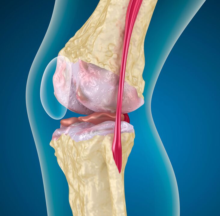
20% of people over the age of 25 are at risk of developing deforming osteoarthritis of the knee. The knee joint operates in an axial compression mode, therefore its articular surfaces are exposed to constant stress and are subject to degenerative changes in the hyaline cartilage.
%20and%20affected%20by%20arthrosis%20(left).jpg)
Prevalence
The pathology of the degenerative-dystrophic articular cartilage involving the bone tissue, joint bag, ligaments and muscles is called arthrosis, which deforms in the process. The terminology has synonyms:
- osteoarthritis;
- osteoarthritis;
- degenerative arthritis;
- arthrosis;
- hypertrophic arthritis;
In terms of frequency, the injury to the knee occurs immediately after the hip joint, thus a stable term was formed: "gonarthrosis of the knee joint". The dependence of the frequency of the disease on age was studied:
| They are 26-44 years old | 5% of adults |
| They are 45-59 years old | 16. 70% |
| They are 60-69 years old | 12. 10% |
| 70 years and older | eleven% |
Quantitatively, representatives of the fair sex predominate in all age groups. In them, knee arthrosis occurs 1. 2-1. 4 times more often than in men.
In the field of permanent disability, deforming arthrosis of the knee joint accounts for nearly 30% of the causes of disability related to joint pathology.
Classification of gonarthrosis
For developmental reasons, the disease is divided into two large groups: primary and secondary. The primary arises without any visible prerequisites. The secondary condition is preceded (or accompanied) by provoking factors:
- biomechanical disorders: injuries, excessive loads, developmental disorders (dysplasia), skeletal pathology (spinal curvature, flat feet), obesity;
- inflammatory processes: aseptic or infectious arthritis, frequent hemarthrosis in hemophilia;
- metabolic diseases: gout, hemochromatosis, Paget's disease;
- endocrine gland disorders: acromegaly, diabetes mellitus, parathyroid gland disorders;
- violation of adequate blood supply: varicose veins and post-thrombophlebitic syndrome, obliterating endarteritis, atherosclerosis of lower limb vessels;
In medical practice, classification according to the severity of pathological changes is more useful. The assessment is based on X-ray examinations. The most popular clinical and radiological classification.
I put it on stage
The image shows a slight narrowing of the interarticular space (the comparison is with a healthy joint), the beginning of sclerosis of the pericartilaginous bone tissue. Clinically - the pain occurs during walking or immediately after standing for a long time. Going up the stairs is more pronounced. It will pass peacefully. Grade 1 gonarthrosis does not significantly affect the function of the joint.
Section II
The joint gap is 2-3 times narrower than normal. Sclerosis is more pronounced, osteophytes are found (spiky growths of bone tissue at the edges of the joint space and along the condyles). The pain is moderate, there are signs of muscle hypotrophy and lameness. The deformation of the knee can be seen in the frontal axis. Grade 2 gonarthrosis leads to noticeable limitation of joint mobility.
Section III
Sclerosis of cartilaginous elements, deformation of joint surfaces. There are areas of subchondral necrosis and local osteoporosis. Cysts in adjacent bone tissue. The joint space is critically narrowed, sometimes not defined. Significant osteophytes. Atrophy of the muscles of the thigh and lower leg, the joint is unstable, there is pronounced deformity. Movement of the knee is sometimes impossible, a contracture develops. During movement - severe pain, lameness.
This classification approach is convenient, as it allows the evaluation of clinical manifestations in relation to organic changes. It provides an opportunity to choose a more effective treatment based on a comprehensive assessment of the condition of the joint.
Development mechanism
The pathogenesis of any arthrosis goes through three stages:
- Damage to cartilage microstructures. As a result of any damaging factor, high molecular weight compounds lose their strength and become enriched with water molecules. The ability of low molecular weight collagens to assemble into macromolecules is impaired. This leads to a decrease in the strength and durability of hyaline cartilage. Chondroprotectors counteract such phenomena.
- If the provoking factor is not eliminated, the weakening of cartilage components (glycosaminoglycans, proteoglycans) continues. This leads to the activation of recovery processes. Their power reserves aren't particularly high, so this stage moves quickly to the next.
- Disruption of compensatory mechanisms leads to the gradual destruction of articular cartilage and the destruction of its cells - chondrocytes. Cartilage cracks extend to the underlying bone. The degree of detachment of cartilaginous components increases, their defibrillation occurs, which leads to thinning of the hyaline membrane.
On the side of the bone, with deforming arthrosis of the knee joint, thickening (sclerosis), cysts and areas of uneven bone density appear. Therefore, the deformation of the joint surfaces and the instability of the joint develop.
Diagnostics
The diagnosis is based on the totality of data obtained as a result of a survey (anamnesis), medical examination and instrumental research methods. The latter include radiographic examinations (CT, MRI), radioisotope (scintigraphy), and arthroscopy.
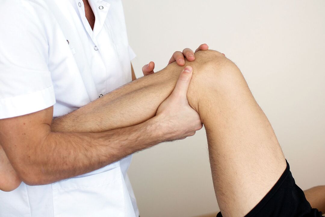
Objective examination
It includes the clarification of the patient's life history, the circumstances leading to the development of gonarthrosis of the knee joint, the collection of complaints and the examination. During the process, the presence of provoking factors and the extent of their impact on the development of the disease are clarified.
At this stage it is important to know the condition of the second knee. If you miss bilateral gonarthrosis and focus only on the knee that worries you more, you can make a gross diagnostic error.
For this, functional tests must be performed on two limbs at the same time. Attention should be drawn to pain during active and passive movements, sensitivity to touch, and crepitus (cracking) occurring during stretching and bending. Chronic inflammatory processes lead to the appearance of Becker's cyst - the protrusion of the joint bag into the popliteal cavity, which can also be detected by palpation.
Instrumental methods
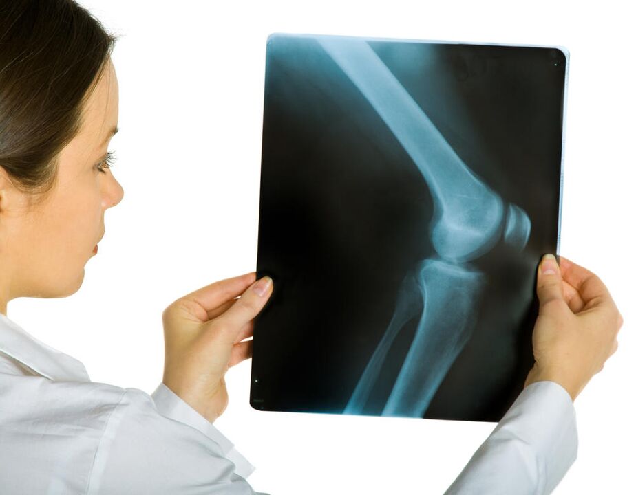
The first is radiography. The image of the knee in two projections enables a preliminary assessment of the condition of the joint and the determination of the stage of the disease. The disadvantage of the method is that the radiological symptoms appear later than the symptoms and morphological changes accompanying arthrosis of the knee joint.
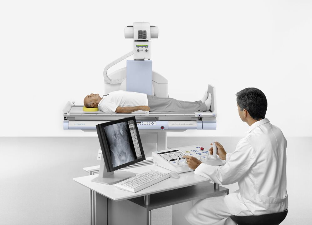
In such cases, MRI (magnetic resonance imaging) helps. The initial stage of degenerative changes in the cartilage and bone structures can be determined, and the condition of the intra-articular ligaments and menisci can be assessed. Scintigraphy of gonarthrosis of the knee joint provides data on the functional state.
Direct examination of the joint cavity is possible with arthroscopy.
To standardize diagnostic data, the American College of Rheumatology proposed the following criteria:
- Age over 50 years.
- Morning stiffness in the joint lasting at least half an hour.
- Crack, determined by movement (active and passive).
If these symptoms are accompanied by osteophytes found on X-rays and pain, there is a high probability of gonarthrosis of the knee joint.
It may happen that the initial stages of the disease are not pronounced, so differential diagnosis should be carried out with other joint pathologies, in which osteoarthritis pathogenetic agents (chondroprotectors) will be ineffective.
All possible measures should be taken in order not to confuse gonarthrosis with the following conditions:
Rheumatoid arthritis |
Morning stiffness lasting more than 30 minutes at an early age, pain at rest and with weaker movement, rheumatic nodules on the skin, simultaneous changes in internal organs, symptoms of poisoning (fever, sweating), C-reactive protein in blood tests. |
Crystal arthritis |
The pain is sharp, at night or in the morning; the skin over the diseased joint is edematous, red, hot; crystals on microscopic examination of the synovial fluid, an increase in uric acid in the blood (in case of gout). |
Spondyloarthropathies |
Arthritis of other unrelated joints (intercostal, lumbar joint); inflammatory processes in tendons; damage to the cornea, skin, mucous membrane. |
In the tenth revision of the International Classification of Diseases (ICD 10), each disease is assigned the "M" index, but with a different numerical code.
These are fundamentally different pathological processes that require a professional approach to diagnosis and skilled treatment.
Therapeutic measures
If there is a disease, it is necessary to find a way to cure arthrosis of the knee joint. And they exist. Help can be provided in several ways.
In the first place are the achievements of traditional medicine, which are based on an in-depth study of the causes and mechanisms of the disease. Medical and surgical methods are used here. Competent treatment requires consistent and complex use of drugs, physiotherapy methods and rehabilitation measures.
The second method is treatment with folk remedies. The effectiveness of these methods is questionable, to say the least. But they are used because the manifestation of the disease can be reduced at home. Folk remedies can only be used as a supplement to drug treatment or as part of complex therapy, the consent of the attending physician is absolutely necessary!
medical help
This type of treatment involves the use of various drugs. Medicines belonging to different groups are used for medicinal effects:
- non-steroidal anti-inflammatory drugs, pain relievers, opiates;
- slow-acting symptomatic drugs (chondroprotectors);
- glucocorticoid hormones;
NSAIDs, rapid pain relievers, opiates
Medicines belonging to this group are designed to eliminate pain. The pain syndrome greatly worsens the life of arthrosis patients, its removal significantly improves the quality of human life. NSAIDs, anilides, non-narcotic and narcotic pain relievers can do this.
A common disadvantage is side effects. These drugs have a negative effect on the kidneys and the protective mechanisms of the gastrointestinal tract. An alternative solution that can reduce harmful manifestations is injections. Intramuscular administration is less damaging to the stomach and speeds up the effect.
Because of the side effects, drugs belonging to this group are prescribed during exacerbations, and careful dose selection is necessary.
The main advantage of NSAIDs is the many forms for local treatment (ointments, gels). It enables the control of the manifestations of the disease at home.
Centrally acting painkillers are prescribed for a short time, in addition to the ineffectiveness of the other two groups. The most popular opiate is prescribed during an exacerbation, more often with bilateral gonarthrosis. These drugs are addictive. You can't pick them up alone!
Symptomatic, slow-acting drugs

The effect of these substances is twofold: they can reduce pain (as NSAIDs) and contribute to the restoration of hyaline cartilage. They are often called chondroprotectors.
The effect develops over several weeks (2-8) and persists for 2-3 months after cancellation.
In addition to chondroitin sulfate and glycosaminoglycans, this group includes hyaluronic acid-based preparations, compounds derived from avocado and soy.
The most studied and popular chondroprotectors (chondroitin sulfate and glycosaminoglycans) are ready-made components of articular cartilage. It is well absorbed in the blood, forming a high concentration in the joint cavity. In order to speed up the effect, the injections can be administered directly into the joint.
Chondroitin sulfate, taken for two years at a dose of 800 mg per day, has been proven to have a stabilizing effect on the joint space in grade 2 gonarthrosis of the knee joint.
Avocado/soy compounds are anti-inflammatory. Due to the inhibition of collagenase (a degrading enzyme), they significantly slow down the destruction of cartilage, increase the synthesis of "own" collagen. In addition, they are very well tolerated.
Hyaluronic acid derivatives are used in the form of intra-articular injections. These foundations, like chondroprotectors, improve the functional state of the knee joint.
The mechanism of action of the different slowly symptomatic drugs is somewhat different, so their combined use is recommended. A high level of safety allows you to take chondroprotectors for a long time without causing tangible damage to the body.
Glucocorticosteroids
The main activity is anti-inflammatory. These funds are prescribed when NSAIDs are ineffective. Tablet forms also damage the lining of the stomach. There are forms for intra-articular injections.
They have many side effects, so you should not abuse hormonal drugs for deforming arthrosis of the knee joint.
Group name |
Advantages |
Mistakes |
|---|---|---|
NSAIDs, pain relievers, opiates |
Quick effect, many forms for local application. |
Side effects, unstable effect, dangerous for age-related patients, addiction occurs. |
Chondroprotectors |
They have a pathogenetic effect, are long-lasting, non-toxic, and there are forms intended for external and intra-articular use. |
Slow development of the effect. |
Hormones |
Rapid action where NSAIDs are insufficient; forms for intra-articular administration. |
Side effects, unstable effect, long-term use is impossible. |
ethnoscience
You can reduce the symptoms of the disease with folk remedies at home. There are many recipes, but there are a few, but:
- no clinical trials have been conducted;
- it is impossible to dose the medicine accurately;
- indications are not clearly defined;
- individual tolerance of folk remedies is not taken into account;
The advantages include a wide therapeutic range, a large selection for external use. Homemade poultices and tinctures and ointments are popular.
The effectiveness of the home treatment can be confirmed by the fact that biologically active substances (gum, bile, herbal infusions) are used for its preparation.
In addition, competent treatment with folk remedies begins with diet and weight loss. This method alone, aimed at reducing the load on the joint, can reverse grade 1 osteoarthritis of the knee joint (the condition is young, sufficient compensatory ability). A healthy diet in itself stimulates the body's ability to regenerate. The diet includes: light hunger, vegetables, freshly squeezed fruit juices. It is advisable to add low-fat jellies and jellies to your diet.
External tools are very diverse. They mainly have an irritating and warming effect. The most studied components are bile, dimethyl sulfoxide and bischofite. Bile must be used medically and must not be extracted from the animal's carcass. Dimethyl sulfoxide is a chemical warfare agent, an analogue of mustard gas. Bischofite is a derivative of oil. This is the original difference.
All three drugs have an anti-inflammatory effect, but should only be used at home after consultation with a doctor. These materials also have contraindications and application characteristics.
We must not forget the placebo effect in the treatment of folk remedies.
The last thing I want to say is that one has one health. You should not completely rely on the apparent simplicity and cheapness of folk remedies. If you have already decided to try them, pay close attention to the painful joint. The progression of the disease against the background of treatment with folk remedies is a reason to reconsider the approach to therapy.
If you are diagnosed with osteoarthritis of the knee joint grade 2 or higher, it is better not to lose from traditional medicine. Or postpone it for a period of remission. Unsatisfactory treatment is an indication for complex surgical intervention.